Previous Research Projects
Depression and Anxiety
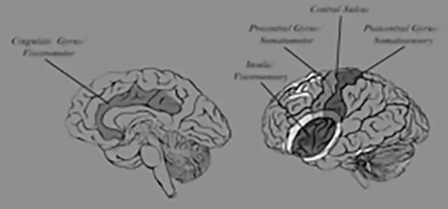
In psychological tasks, such as self-evaluation, the frontal lobe provides regulatory control over emotional responses in limbic regions.
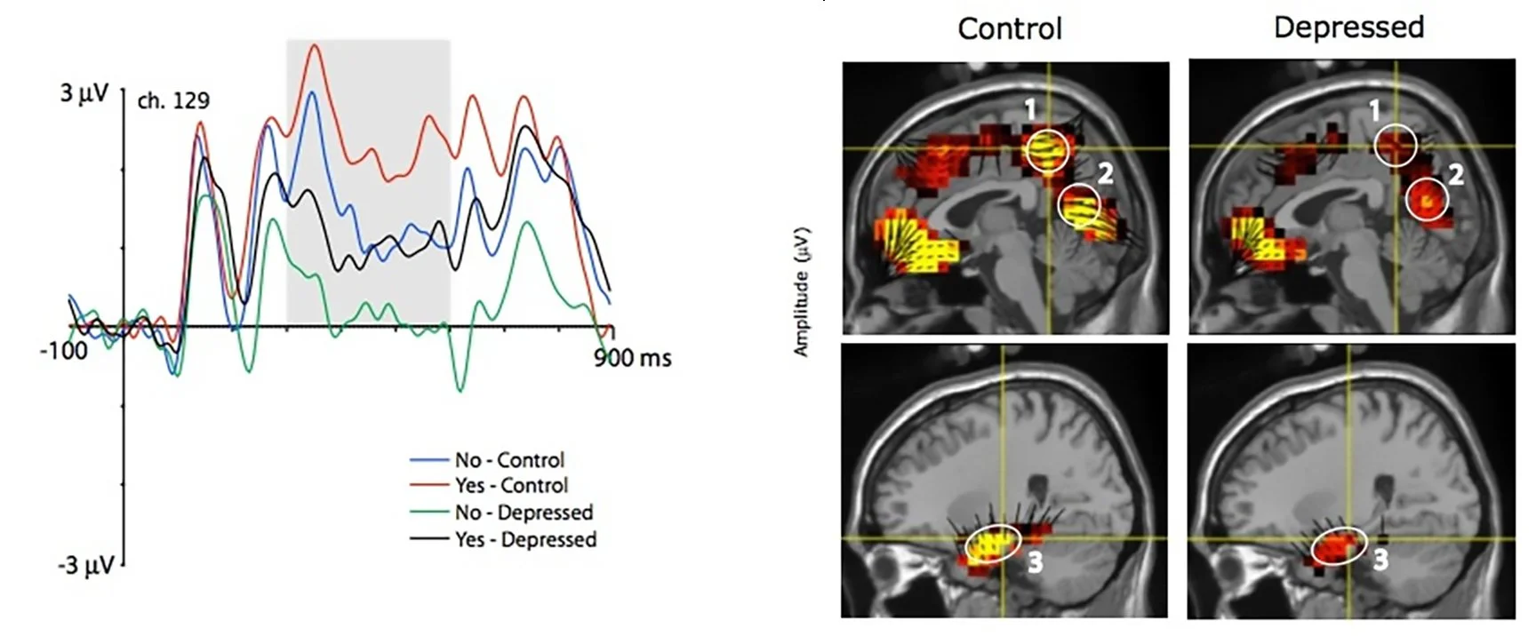
Research in the Brain Electrophysiology Laboratory examines the dynamic changes in frontolimbic activity that discriminates clinically depressed and anxious from normal persons. By understanding these processes, we may be able to guide both biological and cognitive treatments for depression and anxiety.
Chung, G., Tucker, D. M., West, P., Potts, G. F., Liotti, M., Luu, P. et al. (1996). Emotional expectancy: brain electrical activity associated with an emotional bias in interpreting life events. Psychophysiology, 33(3), 218-233.
Luu, P., Collins, P., & Tucker, D. M. (2000). Mood, personality, and self-monitoring: negative affect and emotionality in relation to frontal lobe mechanisms of error monitoring. J Exp Psychol Gen, 129(1), 43-60.
Luu, P. & Tucker, D. M. (2003). Self-Regulation by the Medial Frontal Cortex: Limbic Representation of Motive Set-Points. In M. Beauregard (Ed.), Consciousness, emotional self-regulation and the brain. Amsterdam: John Benjamins.
Tucker, D. M. (2001). Motivated Anatomy: A core-and-shell model of corticolimbic architecture. In G. Gainotti (Ed.), Handbook of Neuropsychology, 2nd Edition, Volume 5: Emotional behavior and its disorders. (pp. 125-160). Amsterdam: Elsevier.
Tucker, D. M., Derryberry, D., & Luu, P. (2000). Anatomy and Physiology of Human Emotion: Vertical Integration of Brainstem, Limbic, and Cortical Systems. In J. Borod (Ed.), Handbook of the Neuropsychology of Emotion. (pp. 56-79). New York: Oxford.
Tucker, D. M., Luu, P., & Derryberry, D. (2005). Love hurts: The evolution of empathic concern through the encephalization of nociceptive capacity. Dev Psychopathol, 17(3), 699-713.
Tucker, D. M., Luu, P., Desmond, R. E., Jr., Hartry-Speiser, A., Davey, C., & Flaisch, T. (2003). Corticolimbic mechanisms in emotional decisions. Emotion, 3(2), 127-149.
Tucker, D. M., Luu, P., Frishkoff, G., Quiring, J., & Poulsen, C. (2003). Frontolimbic response to negative feedback in clinical depression. J Abnorm Psychol, 112(4), 667-678.
Accelerated Learning
In research for the Office of Naval Research's Human Performance, Training, & Education program, BEL scientists are applying neurophysiological theory and methods to understand the neural mechanisms of learning and memory.
From the scientific understanding gained in neurophysiological studies of learning, our goal is to provide direction to Navy trainers on how to optimize rapid and flexible learning and the use of the complex information systems in today's Navy and Marine combat systems. In the simplest case, the result of this research may be improvements in behavioral training, as we understand the underlying brain mechanisms of training.
In a more visionary goal, we may learn to use dEEG at key points during the training process to assess the student's progress in organizing brain activity. The result could be fast and effective coaching on optimal neuropsychological performance.
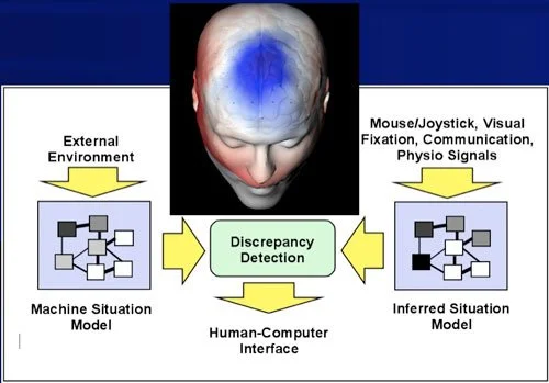
Our current work on brain-computer interfaces is guided by a vision of improved brain-machine communications that was first conveyed in the DARPA Augmented Cognition program, organized by Cmdr. Dylan Schmorrow, ONR.
Luu, P., Tucker, D. M., & Stripling, R. (2007). Neural mechanisms for learning actions in context. Brain Res, 1179, 89-105.
Luu, P., Flaisch, T., & Tucker, D. M. (2000). Medial frontal cortex in action monitoring. J Neurosci, 20(1), 464-469.
Luu, P. & Pederson, S. (2004). The anterior cingulate cortex: regulating actions in context. In M. I. Posner (Ed.), Cognitive neuroscience of attention. (pp. 232-244). New York: Guildford Publications, Inc.
Luu, P., Tucker, D. M., & Makeig, S. (2004). Frontal midline theta and the error-related negativity: neurophysiological mechanisms of action regulation. Clin Neurophysiol, 115(8), 1821-1835.
Poulsen, C., Luu, P., Davey, C., & Tucker, D. M. (2005). Dynamics of task sets: evidence from dense-array event-related potentials. Brain Res Cogn Brain Res, 24(1), 133-154.
Tucker, D. M. & Luu, P. (2006). Adaptive Binding. In H. Zimmer, A. Mecklinger, & U. Lindenberger (Eds.), Binding in Human Memory: A Neurocognitive Approach. (pp. 85-108). New York: Oxford University Press.
Tucker, D. M. & Luu, P. (2007). Neurophysiology of motivated learning: Adaptive mechanisms of cognitive bias in depression. Cognitive Therapy and Research, 31, 189-209.
Training Visual Expertise
In this BEL project funded by DARPA and the National Geospatial-Intelligence Agency (NGA), single-trial measures of dEEG are used to track the expert imagery analyst's visual recognition of military targets in satellite images.
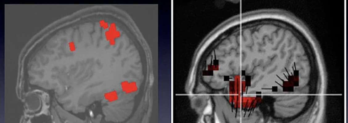
The results showed that dEEG measures were able to reveal the expert's visual response, even when satellite images were presented at rates (10/sec) that were too fast for the analyst to report targets consciously. This finding of brain target recognition was dependent on the ability to characterize the brain's response with millisecond time accuracy and localization of activity to specific brain regions, as shown in the several representations of the dEEG in this image.
An fMRI study of the unique neural responses to detecting targets (left image) showed that detection of military targets engaged not only visual areas (ventral temporal) but memory areas (posterior cingulate). The corresponding dEEG source results (right image) are shown from activity at about 300 ms. In general, the dEEG results showed similar activity in certain regions (including ventral temporal and posterior cingulate), but also showed greater activity in temporal areas than was seen by fMRI.
Whereas comparison with fMRI studies is essential to validate dEEG source results, hemodynamics and electrophysiology provide different views on brain activity, and and this difference remains poorly understood.
Poolman, P., R. M. Frank, P. Luu, S. M. Pederson, and D. M. Tucker (2008). A single-trial analytic framework for EEG analysis and its application to target detection and classification. Neuroimage 42:787-798.


